New method could allow surgeons to assess brain tumor boundaries in ‘real time’
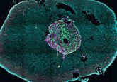
In brain tumor surgery, establishing the boundaries between healthy tissue and tumor cells is key. Typically, pathologists utilize staining methods to identify the location of brain tumors. Chemicals such as hematoxylin and eosin are used to turn different tissue components blue and red, revealing its structure and whether or not there are any tumor cells. However, a definitive diagnosis using this procedure can take up to 24 hr, meaning that neurosurgeons may perform surgery without full knowledge of the cancerous tissue that is present. This incomplete prognosis may mean that a second operation is required, increasing the risk to the...