Brain differences observed in contacts sport athletes
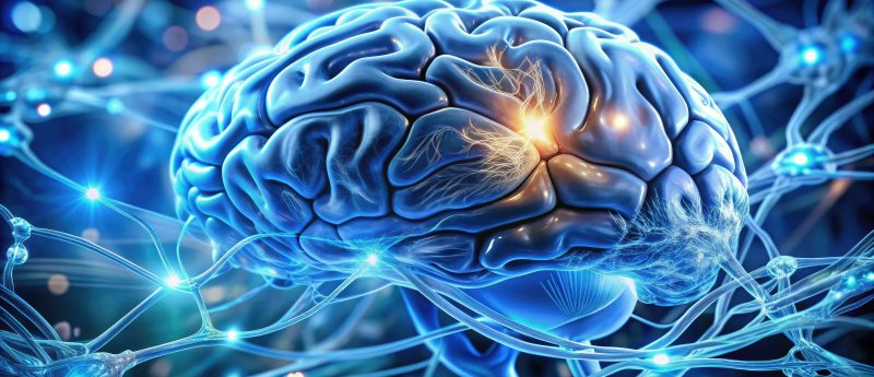
Macro shot of a brain's cerebrospinal fluid filled ventricle, showcasing intricate neural structures and membranes, illuminated by soft blue light in a medical lab setting.
Researchers from St. Michael’s Hospital in Toronto (ON, Canada) have used a combination of MRI techniques to perform preseason brain scans on 65 varsity athletes in a study published in Frontiers in Neurology. The athletes were split into three groups to represent their participation in non-contact, contact or collision sports in the hope that this study would shed light on the brain health of current athletes. Following multivariate analysis, the collision group were observed to display elevated fractional anisotropy and reduced mean diffusivity in white matter compared with other groups. In addition, the collision group demonstrated significant reductions in functional...