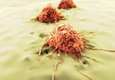
A peek behind the paper – Luis Ibarra on Trojan horse monocyte-mediated delivery of conjugated polymer nanoparticles for improved photodynamic therapy of glioblastoma
In this ‘Peek Behind the Paper’ series, we explore a recent Research Article in the journal Nanomedicine, entitled, ‘Trojan horse monocyte-mediated delivery of conjugated polymer nanoparticles for improved photodynamic therapy of glioblastoma‘. Here, we ask lead author of the study, Luis Ibarra, about what stimulated this interest in using photodynamic therapy (PDT) and nanomedicine to treat glioblastoma. Additionally, we hear more about the advantages of using 3D spheroid models of glioblastoma for testing new therapies.
Please can you introduce yourself and explain what stimulated your interest in using PDT and nanomedicine to treat glioblastoma?
My name is Luis Exequiel Ibarra, I’m a veterinarian with a PhD and postdoctoral degree in science and technology with a focus in nanomedicine. Currently, I’m a researcher from the National Scientific and Technical Research Council (CONICET; Córdoba, Argentina) and also, an Assistant Professor at Universidad Nacional de Río Cuarto (Río Cuarto, Córdoba, Argentina). I belong to a multidisciplinary group with Dr. Rivarola Viviana Alicia, Palacios Rodrigo Emiliano and Chesta Carlos Alberto (all Universidad Nacional de Río Cuarto) with many years of experience in the field of photo-assisted therapies against cancer and in the development of new photoactivatable drugs based on nanotechnology. We are convinced that PDT is a therapeutic modality with many advantages over conventional treatments; and a versatile tool that has allowed its use to be expanded beyond the simple elimination of tumor cells by ROS-generated damage.
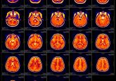
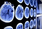
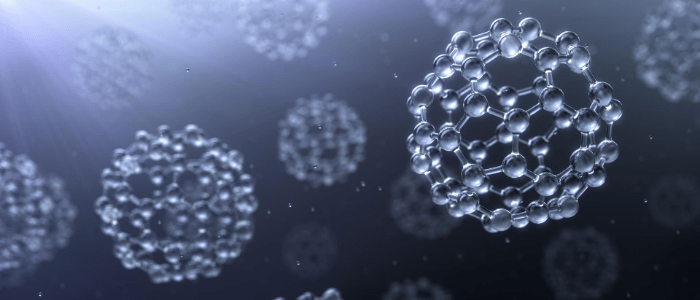
At present, treatments against glioblastoma (GBM) are still in search of surpassing themselves to achieve once and for all the elimination of this aggressive cancer with high mortality rates. In this latter, a particular interest has been placed once again in PDT for the treatment of GBM due to the development of new ways to administer light and the arrival of photosensitizers (PSs) to this preferentially located tumor. Once PDT is applied, toxic effects are not specific to a particular molecular fingerprint of GBM, but rather depend on selective accumulation of the PSs inside tumor cells and perhaps their greater sensitivity to the effects when triggered by light. From PhotoMat, we design and study new nanoparticulate PSs based on conjugated polymers, which have proven to be more effective in collecting photons and transferring excitation energy to PS dopants within the particle for the amplification of ROS generation. This encourages us to continue studying PDT as a main therapy for GBM, or as an adjuvant treatment, to enhance conventional and experimental treatments for the global improvement of GBM management.
What are the drawbacks associated with current PDT approaches?
So far, PDT of GBM is still in clinical trial for classic PSs such as hematoporphyrin derivative, talaporfin sodium, 5-aminolevulinic acid and meta-tetra(hydroxyphenyl) chlorin. All of these are part of the so-called first- and second-generation molecular PSs with relatively modest effectiveness in GBM patients. In Japan, PDT for primary malignant brain tumors was approved by the government for the first time in the world a few years ago, which encourages further research on its therapeutic application. A major problem that PSs face is the lack of antitumor selectivity resulting in high doses managed or low ROS generation quantum yields, which translate perhaps in a selection pressure of resistant tumor cells with subsequent recurrence of the disease after a few months.
In addition, the delivery of light into the brain is another cornerstone, however with the evolution of technology, new devices to provide light have been made available to the PDT community such as interstitial PDT. Now, the third generation of PSs, which can dramatically improve cancer-targeting efficiency by chemical modification, nano-delivery system or ligand conjugation, are extensively studied for clinical development and also evaluated to eradicate GBM tumor cells but remains, in the best case, in a preclinical stage. Finally, we must not forget the tumor biology that this special tumor presents with different heterogeneous regions, with hypoxic areas and tolerance to oxidative stress that can affect the success of PDT.
Could you provide us with an overview of the main findings of your research?
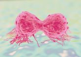
In our work, we studied an alternative method for active targeting of phototherapeutic nanoparticles using the knowledge of GBM tumor biology. A hallmark histological feature of GBM is the infiltrated non-neoplastic immune cell population with a predominant presence of tumor-associated macrophages. This feature could be associated with an inflamed microenvironment, which leads to a favorable environment to the recruitment of leukocytes, mainly circulating monocytes that later differentiate into TAMs. We used monocytes as stealth carriers for the delivery of our conjugated polymer nanoparticles (CPN) into GBM using in vitro and orthotopic models. We observed that monocytes can retain nanoparticles inside without affecting cell function and being able to respond to the chemotactic stimuli released by the tumor. These findings were validated in an orthotopic mouse model using monocytes isolated from bone marrow in a collaboration with the laboratory of Dr Pilar López-Larrubia (High Council of Scientific Researches, Madrid, Spain). Monocytes with a pro-inflammatory profile seems to be more efficient for the Trojan Horse strategy. Finally, CPN-loaded monocytes improved tumor penetration into GBM spheroids compared with non-vectorized nanoparticles and the overall performance of PDT.
In your work, a 3D spheroid model of glioblastoma was used. What are the advantages of 3D tools such as these for testing new glioblastoma therapies?
GBMs are inherently extremely challenging with complex microenvironments resulting in phenotypic heterogeneity that need in vitro models with physiologically relevant parameters like oxygen tension, as well as the changes in the extracellular matrix, which can affect biological properties when investigating GBM cells in vitro and have an impact in therapeutic designs. In addition, 3D spheroid models respond to the 3Rs (i.e., replace, reduce, refine) in the use of animal testing and has advantages over conventional cell culture conditions due to the ability to recreate the complex tumor organization, bioactivity and physiology of GBM. Furthermore, it is possible to evaluate, in a more sophisticated way, the parameters that control the complex penetration depth, biodistribution and tissue diffusion of nanomaterials. Hence, 3D tools provide more realistic microenvironments that allow the evaluation of new therapeutics and mainly, those where the efficacy effect does not depend only on the active drug such as PDT.
What are the next steps your research group will be taking to improve and further test your developed nanoformulation?
The next step for the evaluation of our developed nanoformulation is to determine the safety and anticancer capacity in in vivo models as a first step toward eventual clinical application. On the other hand, we want to understand the role of extracellular vesicles in the delivery strategy of our nanoparticles and if this phenomenon can be enhanced in CPN-carrier cells with metronomic PDT.
Disclaimer
The opinions expressed in this interview are those of the interviewee and do not necessarily reflect the views of Neuro Central or Future Science Group.
You might also like: